
March 15, 2025 · By Alisa Gooding
Trichoscopy Explained: What a Closer Scalp View Can—and Cannot—Tell You
Understand what trichoscopy examines, what to expect during imaging, and why images are one part of a broader hair and scalp assessment.
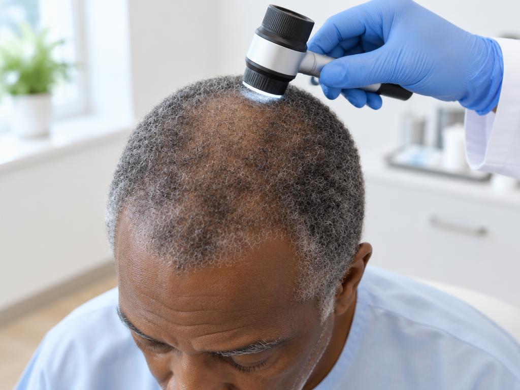
AI-generated educational illustration; not an AGAPE client, clinic photograph, or treatment result.
What does trichoscopy mean?
Trichoscopy is magnified examination of the scalp and hair using a dermatoscope or digital imaging system. It can make features too small to notice with the unaided eye—such as differences in hair-shaft thickness, follicular openings, and surface changes—easier to inspect. In dermatology, it is used as one component of evaluating hair loss, not as a stand-alone answer to every concern.
AGAPE offers its Trichoscopy Suite as high-magnification imaging of scalp and follicles, with images that can provide a baseline for later comparison. The suite is part of a trichology assessment; it is not a substitute for medical diagnosis. A camera can document visible features, but the meaning depends on history, examination, and sometimes evaluation by a dermatologist or other clinician.
What to expect at a session
Before imaging, be ready to explain what has changed and when you first noticed it. Bring a concise timeline of shedding, thinning, breakage, itching, tenderness, styling changes, illness, medication changes, pregnancy, or other relevant events. These details matter because different causes can look similar from a distance. A healthcare professional may also ask about family history and overall health.
Images are generally taken at selected scalp areas and reviewed for visible patterns. Ask what areas are being photographed, how images are stored, and whether comparison photos are planned. If products or fibers are obscuring the scalp, ask the provider whether to arrive with clean, product-free hair; follow the clinic’s specific instructions rather than assuming.
Reading the image in context
Trichoscopy may help a trained evaluator see patterns and document change. It does not by itself prove a specific cause, predict future growth, or establish that a treatment will work. Dermatologists may combine a detailed history and scalp examination with hair testing, laboratory tests, or occasionally a biopsy when appropriate. One image is a snapshot, while follow-up images can help assess whether visible features have changed over time.
AGAPE offers microscopy-based follicle evaluation within trichology care and personalized at-home guidance. That is an assessment and planning service, not a promise of diagnosis or regrowth. If the findings raise concern for a medical scalp condition, a dermatologist is the appropriate clinician to diagnose and treat it.
Make the appointment useful
- Write down when the concern began and whether it is progressing
- Bring a list of medicines and supplements, without stopping anything on your own
- Note painful, rapidly expanding, smooth or inflamed patches and mention them promptly
- Ask what the images show, what they do not show, and whether medical referral is advisable
Come with questions rather than a predetermined diagnosis. Helpful questions include: What was observed? Could breakage be contributing? What would warrant a dermatologist visit? What should be documented at follow-up? A careful assessment explains uncertainty as well as findings. The goal is clearer next steps, not an overconfident reading of a magnified picture.
This article is educational and does not diagnose your symptoms. Seek appropriate medical evaluation for sudden changes, pain, sores, or persistent scalp concerns.